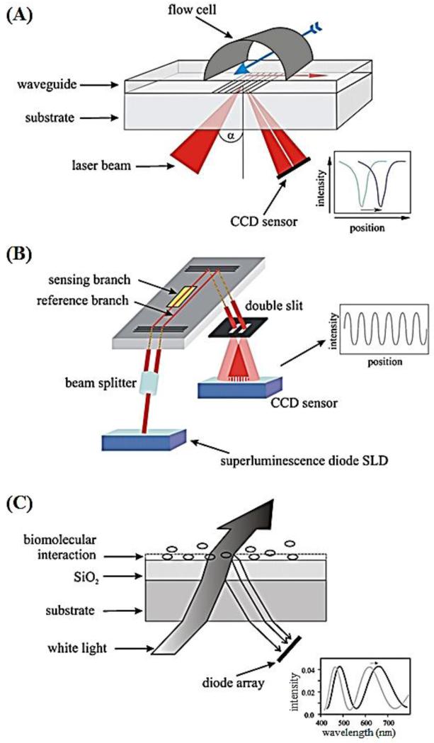
Figure 2.
Schematic of three optical label-free devices for MTB detection (Nagel et al., 2008).
(A) Grating coupler biosensor. A laser beam is shed onto the grating through a waveguide and the reflected light is detected by a charge-coupled device (CCD). The binding of analyte on the sensor surface changes the refractive index, leading to a shift in the coupling angle. (B) Interferometric biosensor. The light (wavelength at 675 nm) is introduced into a waveguide, passed through two branches (one sensing branch and one reference branch) and guided through a double slit. Binding of analyte on the sensor surface causes a change in the interference pattern, which is detected by a CCD sensor. (C) The reflectometric interference spectroscopy (RIfS) biosensor. This sensor is adaptable for detection of white-light interference. When bound to the interface, the analyte causes a shift in the interference pattern in the optical path, which is detected by a diode array.

