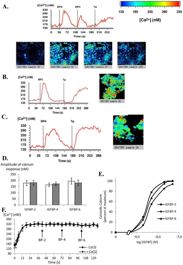
Figure 1. IGFBP-2, -3, -4 and -6 increase intracellular calcium concentrations in MCF-7 cells.
MCF-7 cells cultured on glass coverslips were incubated with Fura-2/AM. A. After background recording for 40 seconds to determine basal intracellular calcium concentrations as described in Methods, cells were incubated with IGFBP-6 (20 nM), then with IGFBP-3 (20 nM) and then with thapsigargin (Tp) (1 µM). The results of a typical experiment are shown in which the whole field (red line) was analysed and intracellular calcium quantified. The slides from left to right show representative views of the cells (a) before addition of IGFBP-6, (b) after addition of IGFBP-6, (c) after addition of IGFBP-3, and (d) after addition of thapsigargin. The results presented are representative of 5 independent experiments. B and C. Same experiments as in panel A excepted that cells were incubated with either 20 nM IGFBP-2 (panel B) or 20 nM IGFBP-4 (panel C). Slides show representative views of the cells after addition of IGFBP-2 or -4, respectively. Results presented are representative of 4 independent experiments. D. Quantitative analysis of the calcium response (maximal calcium response - basal calcium level) obtained for IGFBP-2, -4 and -6 in calcium free (empty bars) or calcium containing (hatched bars) medium. Results are the means ± SEM for three to five independent experiments. E. Dose-response curves for intracellular calcium concentrations were established as described in Materials and Methods. Values are expressed as percentages of the maximal response measured with 50 nM IGFBP-2 and are the mean for two independent experiments. F. Graphs present the average values of the intracellular calcium concentration modulated by addition of 20 nM of IGFBP-2, -4 or -6 following thapsigargin (1 µM) treatment in calcium free or containing medium as indicated.

