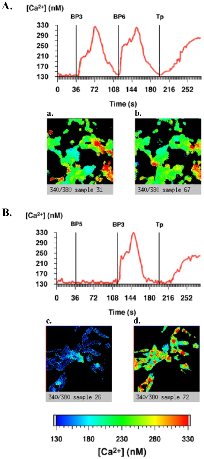
Figure 4. IGFBP-5, but not IGFBP-3 and -6, increases intracellular calcium concentrations via a pertussis toxin-sensitive signaling pathway in C2 cells.
C2 cells cultured on glass coverslips were treated with or without 200 ng.mL-1 pertussis toxin (PTX) for 16 hours at 37°C and incubated with Fura-2/AM. After background recording for 40 seconds to determine basal intracellular calcium concentrations as described in Methods, cells were incubated (A) with IGFBP-3 (20 nM), then IGFBP-6 (20 nM) and then with thapsigargin (1 µM) or incubated (B) with IGFBP-5 (20 nM), then IGFBP-3 (20 nM) and then with thapsigargin (1 µM). The results of a typical experiment are shown in which the whole field (red line) was analysed and intracellular calcium quantified. The slides from left to right show representative views of the cells: panel A (a) after addition of IGFBP-3, (b) after addition of IGFBP-6, panel B: (c) after addition of IGFBP-5, and (d) after addition of IGFBP-3. Results presented are representative of 4 independent experiments.

