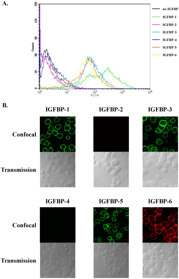
Figure 6. IGFBP-1, -3, -5 and -6, but not IGFBP-2 and -4, associate with C2 cell surface.
A. C2 cells were incubated for 1 hour at 4°C with or without IGFBP-1 to -6 (5 µg.mL-1), fixed and incubated with specific primary antibodies and fluorescent secondary antibodies as indicated in Materials and Methods. Staining profiles were analyzed by FACS and mean fluorescence intensities (MFI) quantified. Data presented are representative of three independent experiments. B. C2 cells growing on coverslips were incubated with IGFBP-1 to -6 (5 µg.mL-1) and then fixed without detergent. Cells were incubated with specific antibodies directed against IGFBP-1 to -6 processed with secondary antibodies stained either with Alexa 488 (IGFBP-1, -3, -4, -5) or Alexa 633 (IGFBP-2, -6). For each incubation condition, upper panel shows confocal fluorescent images and lower panel shows differential interference contrast images. Results are representative of four independent experiments.

