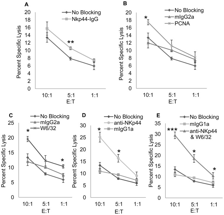
Figure 5. PCNA/HLA I complex interaction with NKp44 inhibits Primary NK Cell Cytotoxic Function.
In order to confirm the results in figure 4, we performed a 51Cr release assay with primary NK cells isolated from Peripheral Blood Mononuclear Cells. Primary NK cells were cultured in 1000 units/ml recombinant human IL-2 for one week, at which point NKp44 expression was confirmed prior to use. DB cells were loaded with 51Cr and incubated with 1 µg/ul of NKp44-Ig to block the entire NKp44 ligand complex (5A) or 0.5 mg/ml anti-PCNA (5B) or anti-HLA I (5C) to individually block PCNA or HLA I interaction with NKp44. DB cells were then incubated with primary NK cells at 10∶1, 5∶1, and 1∶1 effector to target cell ratios for 4 hours at 37°C. Level of killing was compared to DB cells incubated with 0.5 mg/ml mIgG2a isotype antibody or no antibody (No Blocking). In figure 5D, NKp44 was blocked on primary NK cells with 0.5 mg/ml anti-NKp44 or mIgG1 isotype control antibody prior to incubation with DB cells incubated with no antibody. NKp44 on primary NK cells was again blocked with 0.5 mg/ml of anti-NKp44 and incubated with DB cells incubated with 0.5 mg/ml anti-HLA I in figure 5E. Bars ± SD. *p<.05, **p<.01, ***p<.0005, ANOVA.

