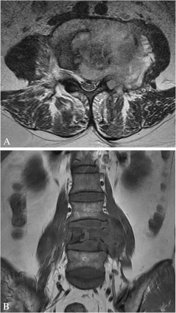
Figure 1.
MRI of the lumbar vertebrae. (A) Transverse section imaging of L4 showed the involvement of left pedicle and transverse process, the paravertebral soft tissue mass and the spinal canal stenosis. (B) Coronal section imaging of lumbar vertebrae showed the osteolytic lesions of vertebral body at L4 level.

