
Fig. 4.
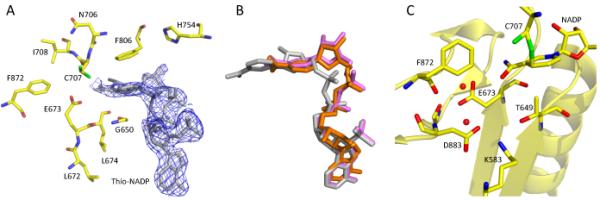
Binding of thio-NADP+ to Ct-FDH. (A) The active site of wild-type Ct-FDH with bound thio-NADP+ (the 2∣FO∣-∣FC∣ electron density map of thio-NADP+ contoured at 1 σ is shown in blue). (B) Overlay of coenzymes bound to ALDHs: grey, NADP+ (extended conformation) bound to wt Ct-FDH; orange, NADPH (contracted conformation) bound to ALDH2 [9]; violet, thio-NADP+ (contracted conformation) bound to wt Ct-FDH. (C) Environment of Glu673 in the complex of Ct-FDH with NADP+ (PDB 2O2Q).