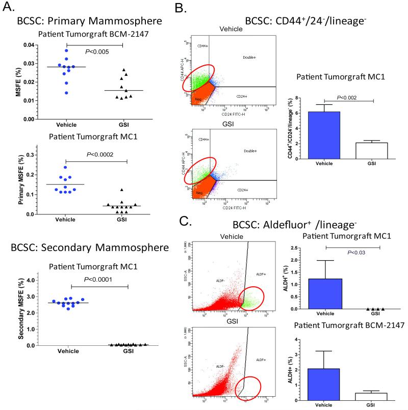
Figure 1. Treatment with GSI reduced the BCSC population in patient-derived breast tumorgrafts.
Mice with tumors were treated for 3 days with GSI (100 mg/kg) or vehicle, and then tumors were collected 72 hours after the last treatment. A. Treatment with GSI reduced mammosphere forming efficiency (MSFE). Tumors were treated in vivo and subsequently plated under MS conditions. To determine secondary MSFE, primary MS were collected, dissociated, and replated under MS conditions. The Mann-Whitney two-tailed t-test was used to calculate p-values. Top figure, each symbol represents an individual BCM-2147 tumor. Middle panel, MC1 tumors cells were pooled for each treatment group. Each symbol represents a well of MS. Bottom panel, MC1 secondary MSFE. B and C. Treatment with GSI reduced BCSC populations according to flow cytometric analysis of BCSC markers, CD44+/CD24− and ALDH+. Treated tumors were dissociated, incubated with antibodies and/or Aldefluor reagents and analyzed by flow cytometry. The Mann-Whitney two-tailed t-test was used to calculate p-values. B. Left column contains representative flow cytometry images. Right column, MC1 CD44/CD24 data (Vehicle n=8, GSI n= 6). C. Left column contains representative flow cytometry images. Top graph MC1 ALDH+ data (Vehicle n=3, GSI n=4). Bottom graph, BCM-2147 ALDH+ data (Vehicle n=9, GSI n=10).

