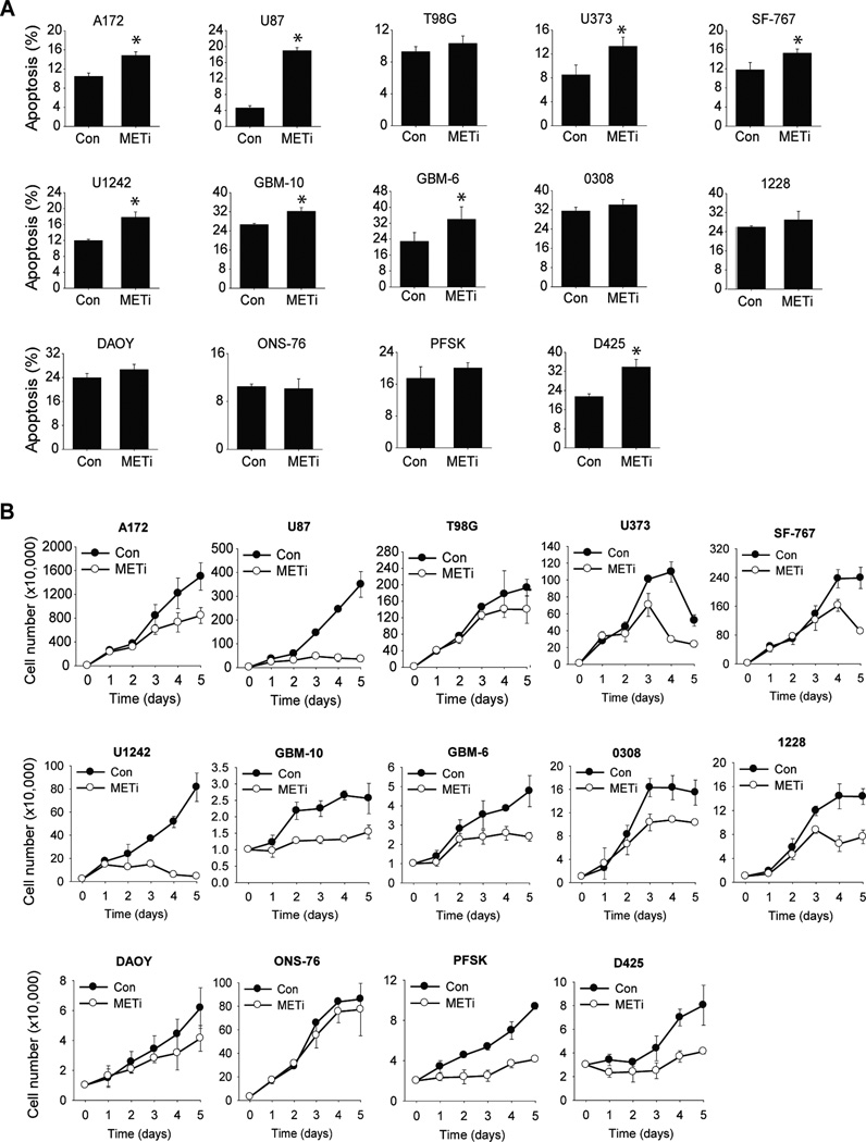
Figure 2. The effects of METi on cell death / apoptosis and proliferation vary between the different brain tumor cells.
A The glioblastoma cell lines U87, A172, T98G, U373, SF-767 and U1242, primary cells GBM-10 and GBM-6, GSCs 0308 and 1228, and the medulloblastoma cell lines D425, ONS-76, DAOY and PFSK were treated with METi or control. The cells were subsequently assessed for apoptosis and cell death by AnnexinV/7AAD flow cytometry and the percentage of dead cells was determined. B The same brain tumor cells as in (A) were treated with METi or control. The cells were subsequently assessed for proliferation by cell counting over a period of 5 days and growth curves were established. All experiments were performed in triplicates and repeated three times.

