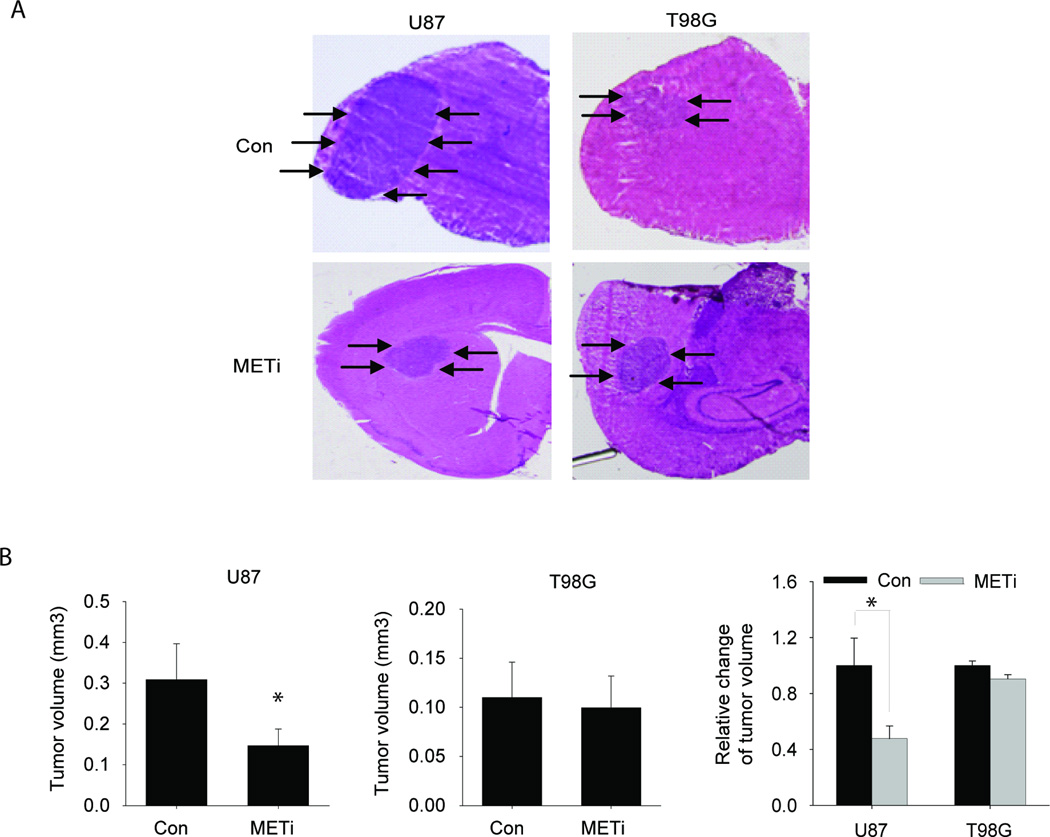
Figure 5. The in vivo anti-tumor effects of METi are significantly greater in high HGF-expressing glioblastoma xenografts than in low-expressing xenografts.
Glioblastoma cells U87 with high HGF-expression and T98G with low HGF-expression were stereotactically implanted in the striatum of immunodeficient mice (n=6 for each treatment group). METi or vehicle control was administered by daily oral gavage starting one week post-tumor implantation. The animals were euthanized 4 weeks after tumor implantation and the volumes of all tumors were measured. The results show that METi significantly inhibited the growth of U87 xenografts but not the growth of T98G xenografts. A Representative brain cross sections with implanted xenografts (arrows). B Quantification of tumor sizes. *, p<0.05.

