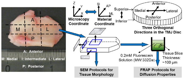
Figure 1.

Schematic of specimen preparation and testing protocols. Each disc was examined in five regions: anterior, intermediate, posterior, lateral and medial. In each region, solute diffusion properties and tissue morphology of these specimens were investigated in three orthogonal planes (i.e., XY, ZX and ZY). The right side discs were tested by FRAP protocols for the diffusion properties and the corresponding left side discs were examined by SEM protocols for tissue morphology.
