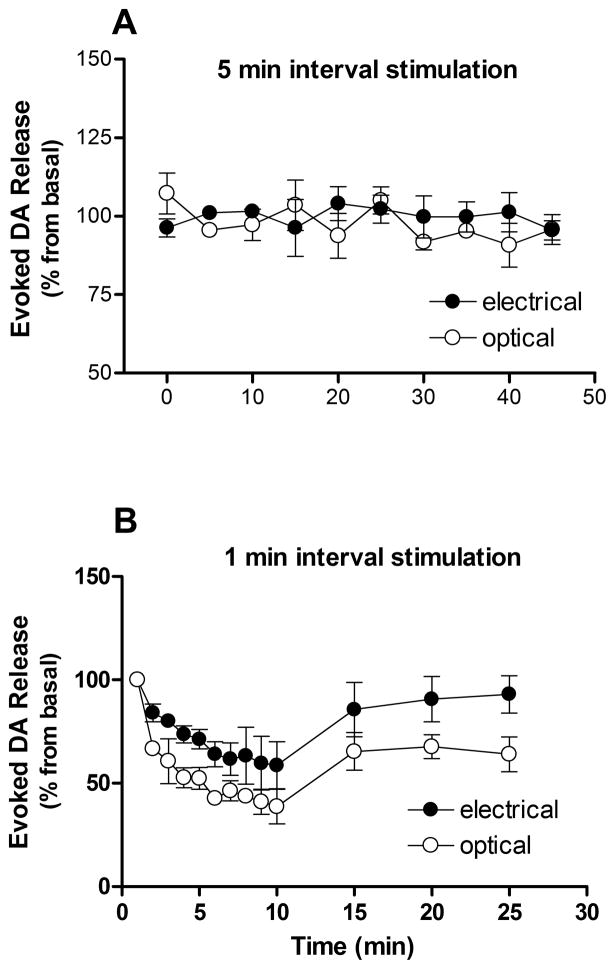
Figure 6. Optically induced dopamine release from striatal slices is stable across multiple stimulations with 5 min intervals.
The stability of light evoked dopamine release (open circles) was compared to electrical stimulation (closed circles). Following every stimulation, there was a significant rise in extracellular dopamine, followed by a decrease back to baseline (Fig. 2). When stimulated at five minute intervals (panel A) there was no difference between stimulation sources and dopamine signal was stable during recording sessions. One minute stimulation intervals resulted in a decline in the amplitude for both optical and electrical stimulation (panel B). Data are means ± SEM of three rats per group, expressed as the percent of dopamine response to the first stimulation.

