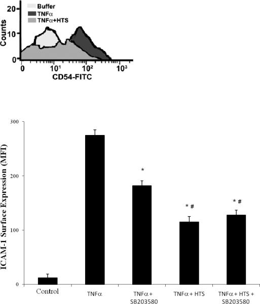
Figure 6.

p38 MAPK inhibition reduces TNFα stimulated ICAM-1 surface expression at normal 140mM [Na+] but does not add to the inhibition provided by HTS (170 mM). HMVECs were pretreated with SB203580 (1μM) in media with either 140 mM or 170 mM [Na+] (300 mOsm/L or 360 mOsm/L) then stimulated with TNFα for 6 hrs. Control is represented, by quiescent cells in 300 mOsm/L media. ICAM-1 surface expression is measured by flow cytometry with the panel showing ICAM-1 positive counts from a representative experimental run. Data are expressed as mean±SEM of 5 experiments. * denotes (p<0.05) difference from TNFα–stimulated HMVECs and # denotes difference from SB203580 pretreated cells, in 300 mOsm/L media (isotonic).
