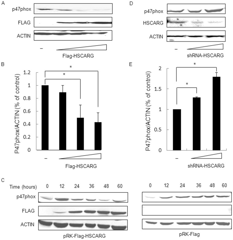
Figure 2. HSCARG inhibits the protein expression of p47phox in a dose/time dependent manner.
(A) Various amounts of HSCARG (0, 0.5, 1.0, 2.0 µg) were transfected into HEK293 cells, and the change of endogenous p47phox protein was examined by western blot analysis using anti-p47phox antibody. (B) Band intensities of p47phox protein level were shown, which were calculated and compared to that of non-transfected cells. (C) The effect of HSCARG on p47phox protein in a time-dependent manner. HEK293 cells were transferred with 2.0 µg HSCARG (left) or control empty vector (right), and the protein levels of endogenous p47phox were examined at different time points (0, 12, 24, 36, 48, 60 h) by western blot analysis using anti-p47phox antibody. (D) Various amounts of shRNA-HSCARG (0, 1.0, 2.0 µg) were transfected into HEK293 cells, and the change of endogenous p47phox protein was examined by western blot analysis using anti-p47phox antibody. (E). Band intensities of p47phox protein level were shown, which were calculated and compared to that of non-transfected cells. Data are presented as mean ± S.E.,n = 3 independent experiments (*p<0.05 vs. control).

