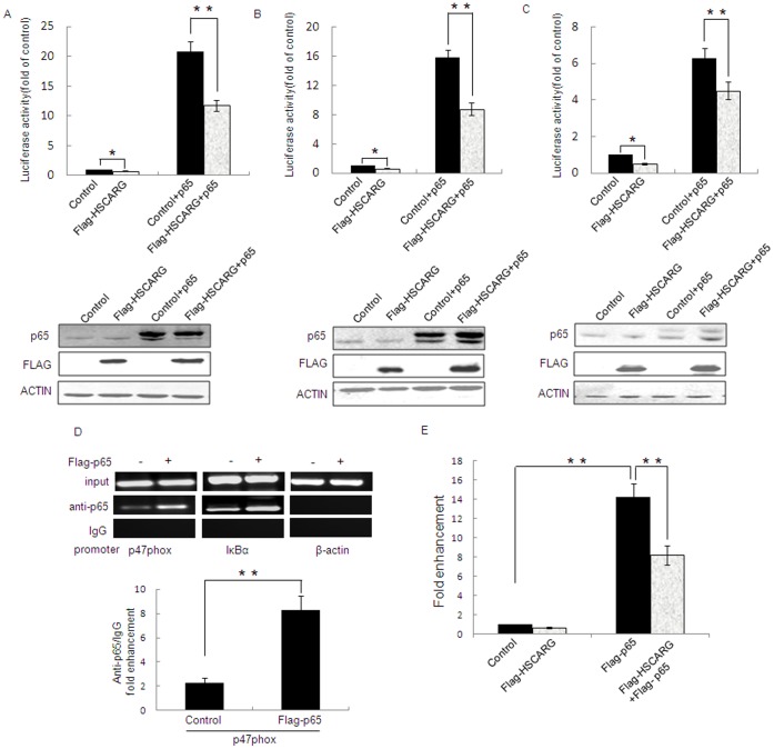
Figure 5. HSCARG reduces p47phox Promoter Activity.
The activity of p47phox promoters -2151 (A), −224 (B), and −86 (C) was examined by luciferase report assay in HEK293 cells transfected with 2 µg HSCARG, pRK-Flag empty vector, pRK-Flag-p65 or pRK-Flag-p65 with pRK-Flag-HSCARG for 48 h, respectively. The expressions of HSCARG and p65 were confirmed by western blot analysis using monoclonal anti-Flag antibody and monoclonal anti-p65 antibody. (D) NF-κB can be recruited to the region from −224 to −1 bp in the p47phox promoter. HEK293 cells were transfected with Flag-p65, pRK-Flag was used as a control. ChIP assay was performed with polyclonal anti-p65 or rabbit anti-IgG (isotype control) antibodies. The immunoprecipitated p47phox promoter (left panel), IκBα promoter (middle panel), or β-actin promoter (right panel) were analyzed by PCR. Quantification of p47phox promoter from ChIP DNA was shown (**p<0.01 vs. control). (E) ChIP assay was quantified by real-time quantitative PCR. HEK293 cells were transfected with p65, HSCARG, and p65 with HSCARG expression plasmids, respectively, pRK-Flag empty vector was used as a control. Values are mean ± S.E. of 3 different experiments (*p<0.05 vs. control, **p<0.01 vs. control).

