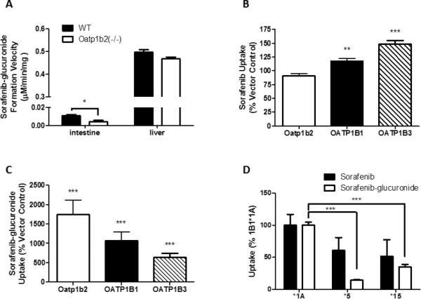
Figure 3. Transport of sorafenib-glucuronide by OATP1B transporters.

A) Ex vivo sorafenib-glucuronide formation was determined in intestine and liver microsomes from wildtype (WT) and Oatp1b2(-/-) mice. Microsomes (1 mg/mL) were incubated with 10 μM sorafenib for 60 min and sorafenib-glucuronide formation velocity was determined. Data represent the mean ± SE of 4 (intestine) or 16 (liver) samples. B, C) HEK293 cells expressing Oatp1b2, OATP1B1, or OATP1B3 were incubated with 10 μM [3H]sorafenib (B) or 10 μM sorafenib-glucuronide (C) for 15 min and intracellular concentrations were determined by liquid scintillation or LC-MS/MS, respectively. Data represent the mean ± SE drug uptake relative to vector control cells from 2 experiments performed with triplicate samples (n = 6). D) T-Rex293 cells expressing OATP1B1*1A (*1A), OATP1B1*5 (*5), or OATP1B1*15 (*15) were incubated with 0.1 μM [3H]sorafenib or 1 μM sorafenib-glucuronide for 15 min. Data represent the mean ± SE drug uptake of 6-15 replicates (*, P<0.05; **, P<0.01; ***, P<0.001).
