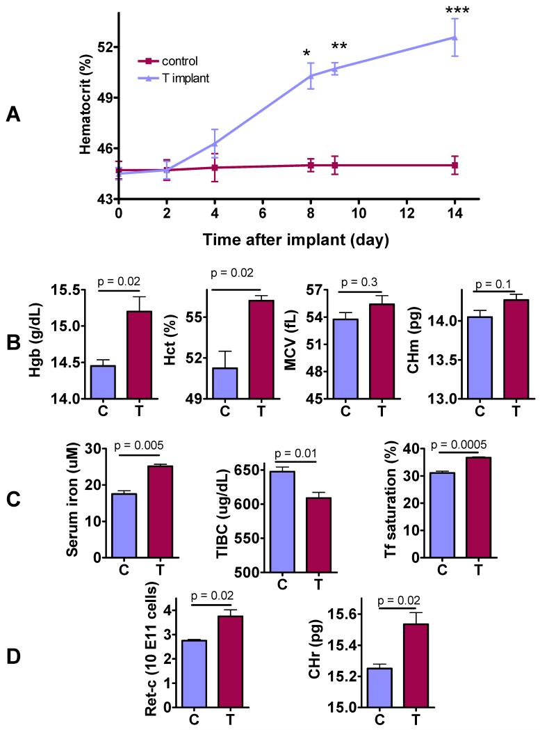
Figure 1. Effects of Testosterone on Red Cell Indices, Reticulocyte Count, Reticulocyte Hemoglobin Ratio, and Circulating Iron Indices.
A: Time course of hematocrit change after subcutaneous insertion of empty or testosterone implants in female mice (*p=0.0002, **p<0.0001, ***p=0.0002 for between-group comparison at each time). The slope of hematocrit change over time was 0.65 for testosterone group (p<0.0001) and 0.02 for control group (p=0.6121).
B: The mice treated with testosterone implants for 2-weeks had significantly higher hemoglobin (Hgb), hematocrit (Hct), and a trend towards higher mean corpuscular hemoglobin (CHm) than mice that received empty implants.
C: Testosterone-treated mice had significantly higher serum iron, lower total iron binding capacity (TIBC), and higher transferrin (Tf) saturation than controls 2 weeks after insertion of either empty (C) or testosterone implants (T).
D. Testosterone-treated mice (T) had significantly higher reticulocyte count (Retic-C) and higher reticulocyte hemoglobin ratio (CHr) than controls (C) after 2 weeks of treatment.
The data are mean±SEM, n=10-20 mice for each measurement.

