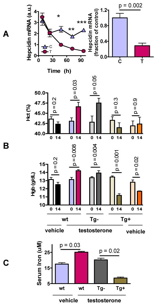
Figure 2. The effects of testosterone on hepcidin mRNA expression and on hematocrit, hemoglobin, and serum iron in wild type and hepcidin transgenic mice.
A. Testosterone suppresses hepatic hepcidin expression. Testosterone administration was associated with a greater decrease in hepatic hepcidin mRNA expression than vehicle after 96h (left panel, *p<0.0001, **p< 0.0001, ***p<0.0001 for between-group comparison at each time) and 2 weeks of treatment (right panel).
B. Hematocrit (Hct) (upper panel) and hemoglobin (Hgb) (lower panel) in wild type (wt) and transgenic female mice that carried either a silent (Tg−) or a constitutively active (Tg+) hepcidin transgene at baseline (day 0) or on day 14 after administration of either empty or testosterone implants. Wt and Tg− mice both displayed higher Hgb and Hct on day 14 compared to baseline in response to testosterone administration, whereas Tg+ mice displayed a slight decrease in Hgb and Hct. Wild-type mice treated with empty implants displayed a small decrease in Hgb and Hct likely due to repeated blood drawing.
C. Serum iron levels in female wild type (wt) and mice carrying silent (Tg−) or constitutively active (Tg+) hepcidin transgene. Testosterone treatment for 2 weeks was associated with significantly higher serum iron levels in wt and Tg− mice than in vehicle-treated controls and in testosterone-treated Tg+ mice. Tg+ mice had significantly lower serum iron than all other groups.

