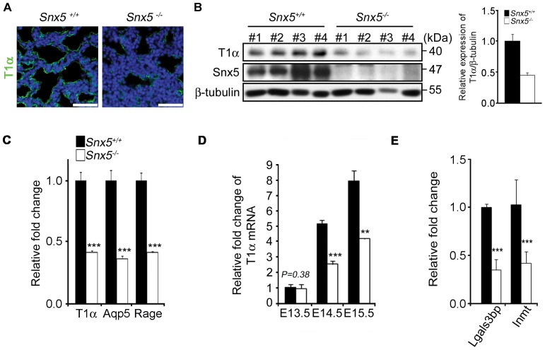
Figure 6. Impaired differentiation of alveolar epithelial type I cells in E18.5 Snx5-/- lungs.
(A) Immunohistochemical analysis of T1α at E18.5. T1α, a marker of alveolar epithelial type I cells, was significantly decreased in Snx5-/- compared to Snx5+/+ lung (n = 5). The nuclei were stained with Hoechst. Scale bars: 50 µm. (B) Decreased protein expression of T1α in E18.5 (n = 4). Densitometric analysis showed that average expression levels of T1α in Snx5-/- lungs were reduced compared to those in Snx5+/+ lungs. (C–E) qRT-PCR analysis of type I cell-related genes from lung tissue. (C) Markers of alveolar epithelial type I cells, i.e., T1α, Aqp5, and Rage, were decreased in Snx5-/- lungs (n ≥ 3). (D) T1α mRNA during the embryonic stage. From E14.5, mRNA expression of T1α was decreased. (E) Decreased expression levels of Lgals3bp and Inmt (n ≥ 3). **, P < 0.01, ***, P < 0.001 using the Student’s t-test.

