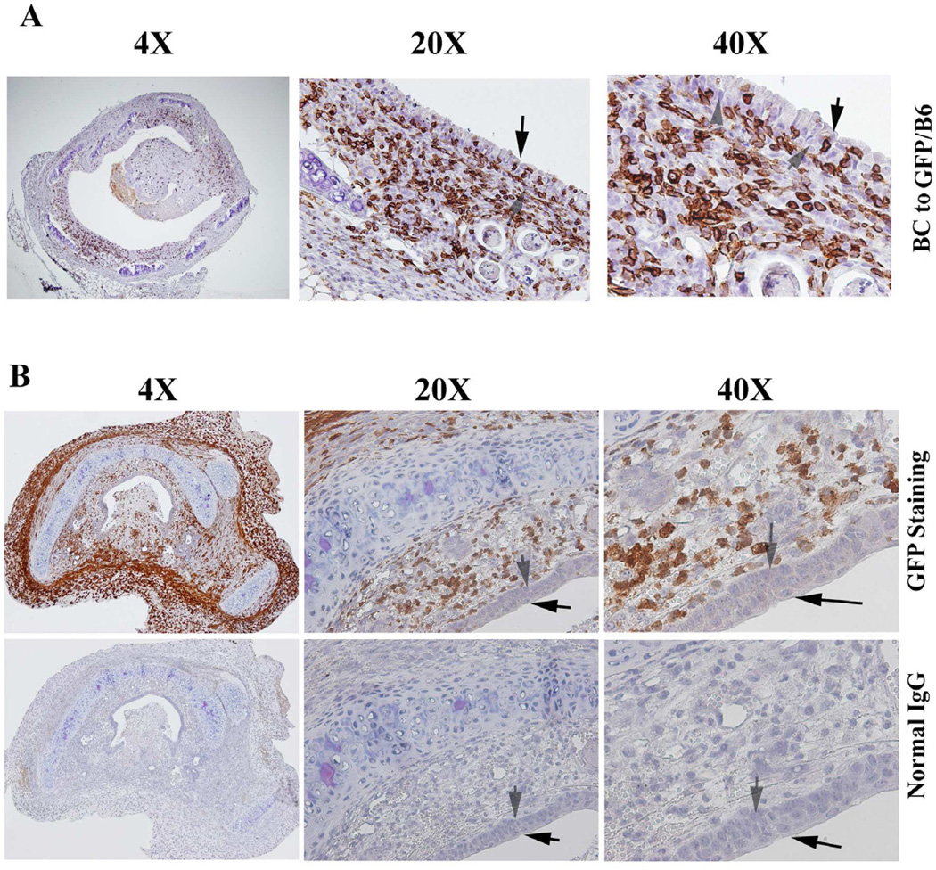
Figure 4.
Immunohistochemical staining of CD3+ T-cells in (A) the allografts of Balb/c to GFP/C57BL/6, and in (B) isograft of C57BL/6 to C57BL/6. Cells stained brown indicate CD3+ T-cell infiltration. All sections were counterstained lightly with hematoxylin for viewing negatively stained cells. The slides were stained with anti- CD3 antibody. Normal IgG was used as control. The magnifications were indicated in the pictures.

