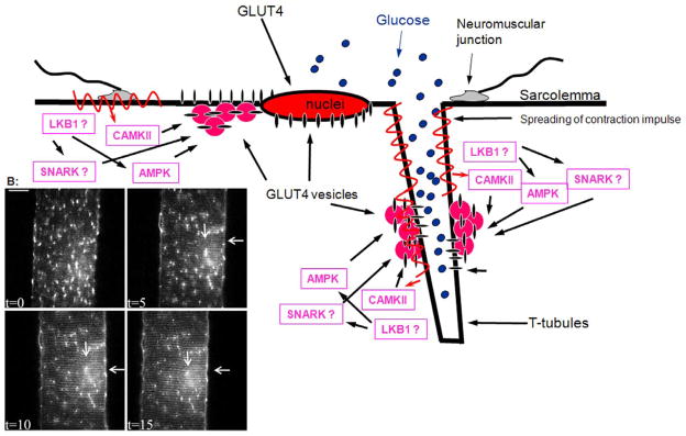
Fig 4. Contraction mediated GLUT4 trafficking in mature muscle.

(A) Several parallel signalling pathways lead to contraction mediated GLUT4 translocation. Intravital imaging has not found any indication of differential compartmentalization in regard to calcium activated calmodulin dependent kinase (CAMKII) - or AMP activated kinase (AMPK) - activation induced GLUT4 translocation. However, any local differences in Serine/threonine kinase 11 (LKB1) or the AMPK related kinase sucrose non-fermenting AMPK related kinase (SNARK) signalling have not yet been studied. Therefore potential compartmentalization of these signals remains to be elucidated. (B) Intravital imaging of contraction mediated GLUT4-EGFP translocation in a part of a muscle fiber in situ in a living mouse (for further details see (19)). At t=0, GLUT4-EGFP is localized to larger and smaller vesicular depots throughout the muscle fiber and around the lateralized nuclei. Immediately after t=0 in situ contractions were initiated for 3×5 min periods (for further details see (19)). After each 5 min period, an image was taken at t=5, t=10 and t=15. It is seen that GLUT4-EGFP is gradually being inserted into the sarcolemma indicated by staining along the plasma membrane (horizontal arrow). Simultaneously a striated pattern is gradually emerging indicating GLUT4-EGFP translocation to t-tubules (vertical arrow). This indicates that translocation to both membrane surfaces takes place with similar kinetics (for quantification see (19). In addition, the basal GLUT4-EGFP depots have gradually been depleted with contractions. Bar = 20 μm.
