
Figure 4. Interactions of Nutlin-3 with MDM2 and MDM4.
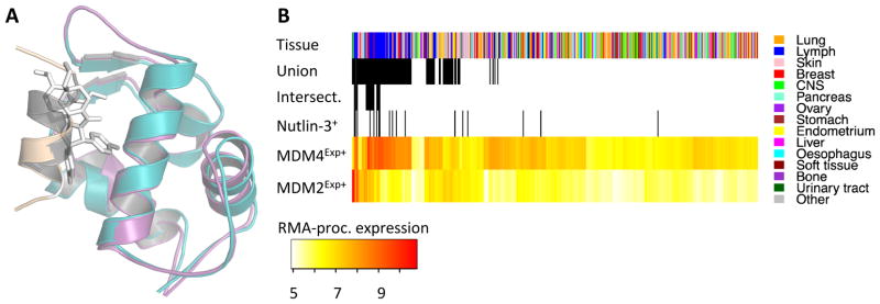
Panel A shows superimposed crystal structures of p53-bound MDM2 (MDM2, teal; p53, beige) and MDM2 binding a nutlin-3 analog (stick representation; MDM2 removed for clarity), and p53-bound MDM4 (MDM4, magenta; p53, gray). Distribution of selected predictive features of nutlin-3 response (B). The superscript Exp+ delineates overexpression (RMA processed) in all panels and Nutlin-3+ delineates samples sensitive nutlin-3. Intersect is the intersection of MDM4Exp+, MDM2Exp+, and Nutlin-3+, and Union is the union of those three features. Lymph is lymphoid and heamatopoietic tissue.