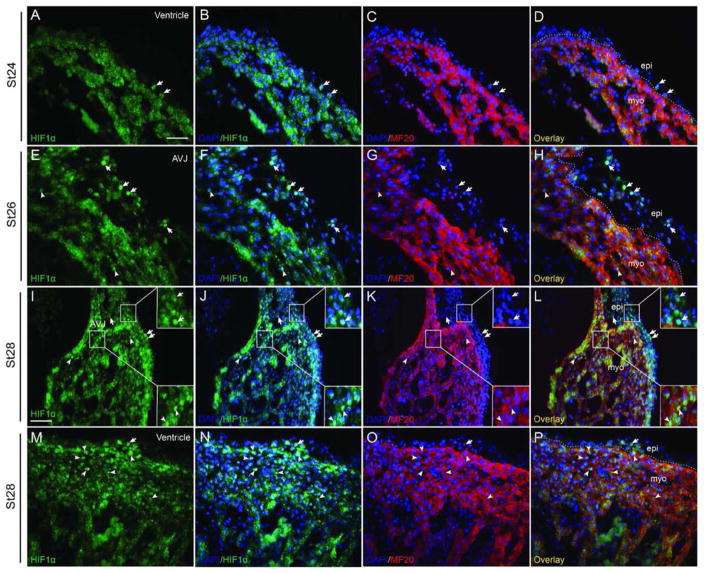
Fig 1. HIF-1α expression in the epicardium and subepicardium.
Costaining for HIF-1α and the myocardium marker MF20 reveals the HIF-1α staining in the epicardium and subepicardium during stage HH24-28. HIF-1α expression was observed in a few ventricular epicardial cells at stage HH24 (A–D) and stage HH28 (M–P). (E–L) HIF-1α is especially intense in the epicardium and subepicardium at the AVJ at stage HH26 (E–H) and continued to be nuclear-localized in this area to stage HH28 (I–L). HIF-1α was also detected in myocardial cells (E–P). Dashed lines delineate the border between the myocardium and the epicardium/subepicardium. Arrows point to HIF-1α positive epicardial or subepicardial cells and arrowheads point to HIF-1α positive myocardial cells. Bar = 20 μm for A–H and M–P, Bar = 40 μm for I–L.

