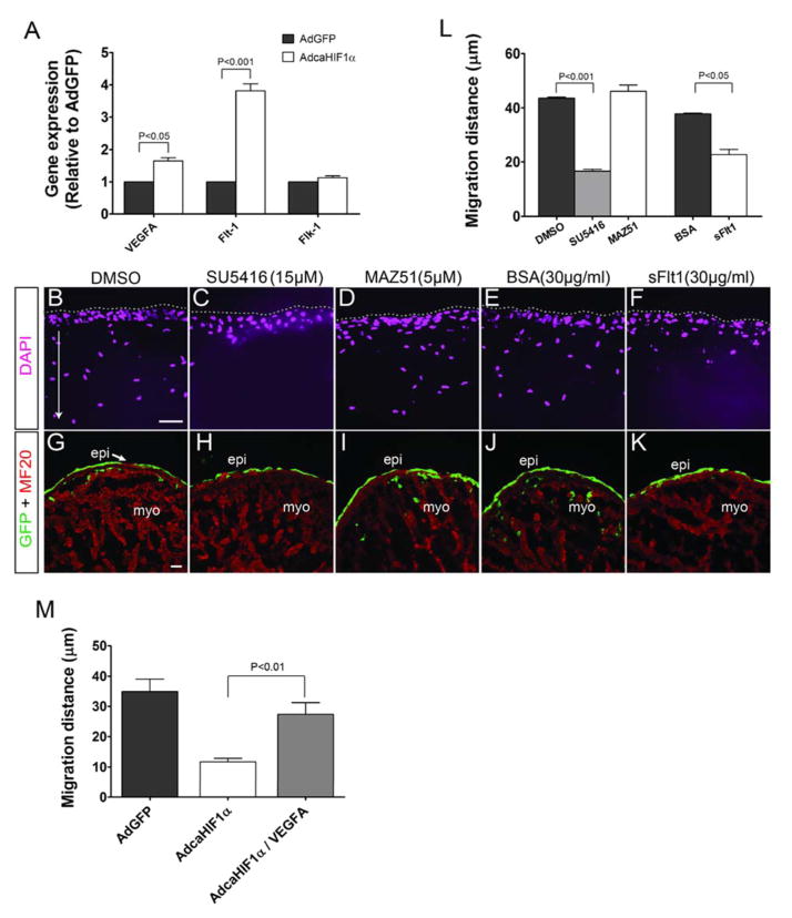
Fig 7. Increased levels of Flt-1 that inhibits Flk-1 signaling could be the mechanism for the inhibition of EPDC migration into the myocardium.
(A) Expression of important factors involved in the VEGF signaling pathway was evaluated using qRT-PCR analysis in chicken epicardial cells, n=3. The level of Flt-1 mRNA expression was much higher in AdcaHIF1α transfected cells compared with the AdGFP transfected cells. (B–K) Data from in vitro collagen gel assays (B–F) or heart explant cultures (G–K) showed that chicken epicardial cells invaded efficiently in the vehicle control DMSO (B, G), with the BSA alone control (E, J), and even with the addition of the VEGFR3 specific inhibitor MAZ51 (D, I). In contrast, the invasion of epicardial cells into gels or the myocardium was inhibited by the Flk-1 specific inhibitor SU5416 (C, H), or Fc-sFlt1 which was utilized to trap VEGF ligands (F, K). Treatments are indicated at the top of the panel. Scale bars represent 20 μm for B–F and 25 μm for G–K. (L) Quantification of average migration distances of epicardial cells in collagen gel assay (Normalized to comparable control group: DMSO or BSA group). (M) Collagen gel assay indicated that inhibition of EPDC migration was partially rescued by VEGFA (50 ng/ml) treatment.

