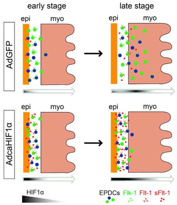
Fig 8. Model of how hypoxia regulates EPDCs invasion into the ventricular myocardium.
In AdGFP-injected controls, HIF-1α is expressed in some epicardial cells at an early stage (stage HH24) and its expression in the myocardium increases at later stages (i.e., stage HH28). This pattern guides EPDCs invasion through the subepicardium and myocardium. However, disruption of the normal pattern of HIF expression with a sustained expression in the epicardium mediated by adenovirus caHIF1α results in the up-regulation of expression of Flt-1 (possibly for both membrane and soluble forms), which leads to the inhibition of EPDC migration into the myocardium.

