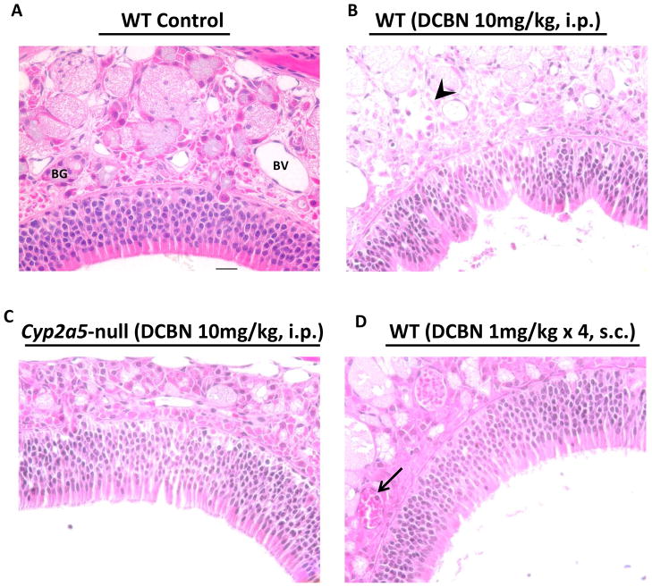
Fig 6. Histopathological analysis of nasal mucosal damages caused by DCBN treatment in WT and CYP2A5-null mice.
Two- to three-month old mice were treated with a single i.p. injection of DCBN (at 10 mg/kg), or four consecutive subcutaneous injections of DCBN (at 1 mg/kg, and with 1.5-h intervals), or with the vehicle alone. Tissues were obtained for histopathological examination at 24 h after the last treatment. Representative H&E-stained cross sections of the dorsal medial region of the nasal cavity, obtained at the level 3,36 are shown. (A) Vehicle-treated mice had normal lamina propria with intact Bowman’s glands (BG) and clear blood vessels (BV). The lamina propria is covered by a pseudostratified olfactory neuroepithelium of normal thickness. (B) DCBN-treated WT mice (10 mg/kg, i.p.) showed moderate injury to the nasal mucosa, evident by deformed epithelium still attached to the basement membrane, and degenerated BG in the lamina propria (arrowhead). (C) DCBN-treated Cyp2a5-null mice (10 mg/kg, i.p.) showed essentially normal olfactory epithelium and BG. (D) DCBN-treated WT mice (4 × 1 mg/kg, s.c.) showed essentially normal morphology, except for aggregation of red blood cells in BV and occasional presence of neutrophils (arrow). Scale bar: 20 μm.

