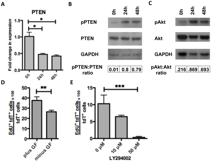
Figure 4. Proliferation of cells of neural crest origin is regulated by P13K/Akt signaling pathway via loss of PTEN inhibition.
We observed a two-fold reduction in PTEN mRNA expression at 24 h and 28 h compared to baseline (0 h) by qRT-PCR (A). Although total PTEN protein levels did not differ there was an 80-fold elevation in the ratio of phospho-PTEN:total PTEN (where phospho-PTEN represents inactive state) at 24 h and 48 h on western blot analysis (B). This was accompanied by a 4-fold and 3-fold increase in the ratio of phospho-Akt:total Akt at 24 h and 48 h, respectively, on pixel intensity of western blot analysis (C). The presence of growth factors (bFGF, EGF, and GDNF) in media caused a modest increase in proliferation at 48 h (D). When organotypic cultures were treated with the PI3K inhibitor LY294002 (without growth factors) for 30 h, a reduction in proliferation was seen at both 10 µM and 50 µM concentrations (E). * = p<0.005 by one-way ANOVA with Bonferroni’s multiple comparison, ** = p<.05 by t-test, *** = p<.01 by one-way ANOVA with Bonferroni’s multiple comparison. Error bars indicate SEM.

