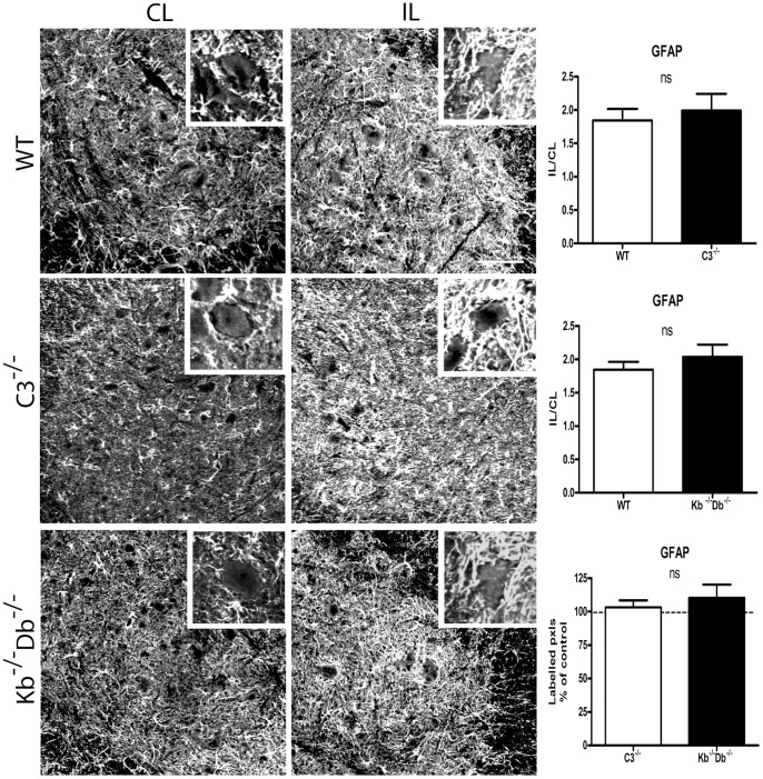
Figure 2. No difference in astrocyte activation as measured by GFAP IR between WT, C3−/− and Kb−/−Db−/− mice one week after sciatic nerve lesion.
Immunoreactivity for GFAP was measured in the sciatic motoneuron pool for WT mice (first row), C3−/− (second row) and Kb−/−Db−/− mice (third row). Ipsilateral (IL, second column) to contralateral (CL, first column) ratio of semiquantative measurements was quantified in the third column. In WT mice, GFAP IR IL/CL ratio was increased to 1.99 and 1.85 of the value on the control side in the two studied sets of experiment. In C3−/− mice the corresponding increase in IL/CL ratio was 1.84, and in Kb−/−Db−/− mice 2.04. Insets showing 50× magnification micrographs of GFAP IR around individual motoneurons. Six animals were studied in each group. Error bars indicate SEM, unpaired t-test. Scale bar = 50 µm.

