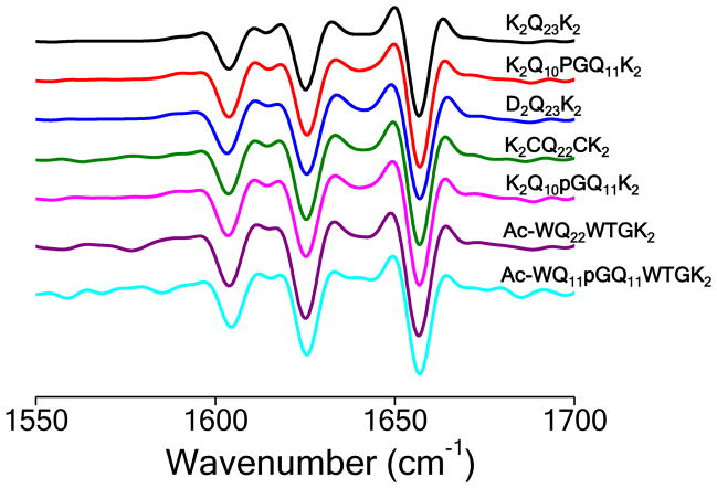
Figure 6.
Secondary structures of amyloid fibrils. Second derivative Fourier transform infrared spectra of isolated amyloid fibrils. The three major bands in these spectra are typical for polyQ aggregates and are assigned to NH2 deformations in the Gln side chains (~ 1605 cm−1), β-sheet (1625–30 cm−1), and C = O stretching in the Gln side chains (1655–1660 cm−1) 48.

