
Figure 8.
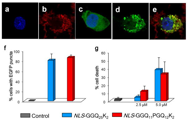
Cell activities of aggregates. (a–e) Confocal fluorescence microscope images of PC12 cells stably transfected to express a Huntingtin exon-1 fragment containing a Q25 repeat and fused to green fluorescent protein, 24 hrs after induction with 1 μM ponasterone with (a,b,d,e) or without (c) treatment with tagged amyloid fibrils. (a) Blue only, Hoechst 33342 (Invitrogen) stained nuclei; (b) red only, Cy5-labelled NLS-GGQ11PGQ12CKK amyloid; (c) blue-stained nucleus and diffuse green showing non-aggregated exon1 in cells not treated with exogenous aggregates; (d) green only, showing aggregate formation from exon1 protein in cells treated with exogenous aggregates; (e) merge colors in aggregate-treated cells showing that aggregates outside the cell remain red, while aggregates within the exon1-producing cell are mostly overlapping red + green. (f) cellular recruitment, as percent PC12 cells with EGFP inclusions, after 16 hrs of expressed (1 μM ponasterone induction) Q25 exon1-EGFP by polyQ fibrils internalized from a 2.5 μM (monomer equivalent) suspension in the growth medium; control data is for similarly induced cells not treated with aggregates. (g) cell death, assessed after 24 hrs by LDH release assay, in PC12 cells treated with different concentrations of aggregates in the growth medium; control data is for cells grown without aggregates.