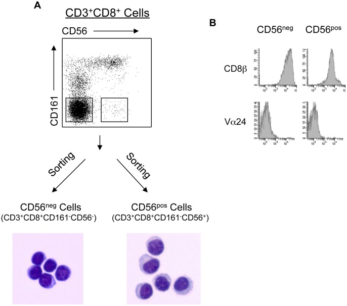
Figure 4. CD161−CD56+ CD8 Treg are CD8αβ+Vα24− cells with abundant cytoplasm.
(A) Morphology of CD161−CD56+ CD8 Treg. PBMC were stained as described in Materials and Methods. CD56pos (CD3+CD8+CD161−CD56+) CD8 Treg and CD56neg (CD3+CD8+CD161−CD56−) CD8 Tcon were purified by cell sorting. Isolated CD56pos and CD56neg cells were stained with Wright-Giemsa stain and visualized using a bright-field microscope. Photos are from one representative experiment of six. (B) Cell surface expression analysis of CD8β and Vα24 on CD56pos and CD56neg cells. CD56pos (CD8 Treg) and CD56neg (CD8 Tcon) were purified by fluorescence-activated cell sorting. Both subsets were CD161 negative. Sorted cells were analyzed for expressions of CD8β and Vα24 as described in Materials and Methods. Data are from one of four representative healthy donors.

