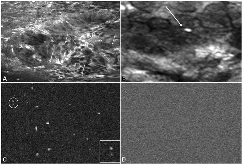
Figure 2. In vivo visualization of intramucosal bacteria within the colonic mucosa in C. difficile colitis by confocal laser endomicroscopy.
Fluorescence confocal image below the surface of the colonic mucosa after topical application of acriflavine hydrochloride identified single bacteria (Panel A, arrows). At 10,000 fold digital magnification the rod-like appearance of bacteria (arrow) in the colonic mucosa became visible (Panel B). Panel C shows ex vivo imaging of pure cultured C. difficile at 1000-fold magnification and 10,000 fold magnification (insert in lower right quadrant) after staining with acriflavine hydrochloride. In contrast, after application of fluorescein no bacteria were visible by confocal imaging (Panel D).

