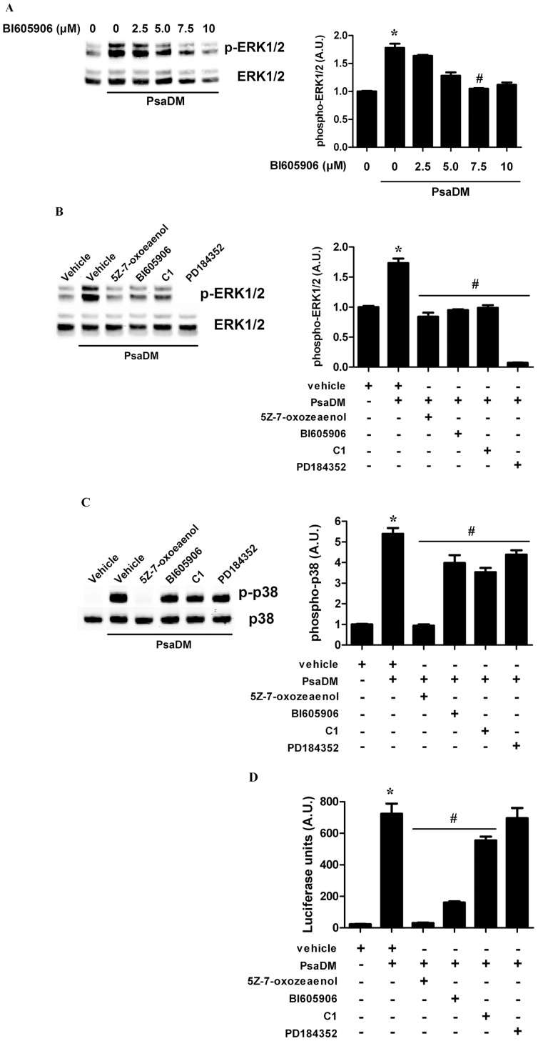
Figure 3. The protein kinases TAK1 and IKKβ are required for MKK1/MKK2-ERK1/ERK2 activation in BEAS-2B AECs stimulated with PsaDM. A.
BEAS-2B AECs were left untreated or pre-treated 1 hour with increasing doses of the IKKβ inhibitor BI605906 (0–10 µM) and exposed to 5 µg/ml PsaDM for 15 minutes. ERK1/ERK2 phosphorylation was detected as in Fig. 1 . B–C. BEAS-2B AECs were pre-treated for 1 hour with vehicle, TAK1 inhibitor 5Z-7-oxozeaenol (0.25 µM), IKKβ inhibitor BI605906 (7.5 µM), C1 (2 µM) or PD184352 (2 µM) and stimulated with 5 µg/ml PsaDM for 15 minutes. Lysates were immunoblotted for ERK1/ERK2 (B) and p38α (C). D. BEAS-2B AECs stably transfected with pGL4.28-NF-κB were left untreated or pre-treated for 1 hour with vehicle, TAK1 inhibitor 5Z-7-oxozeaenol (0.25 µM), IKKβ inhibitor BI605906 (7.5 µM), C1 (2 µM) or PD184352 (2 µM) and stimulated with 5 µg/ml PsaDM for 2 hours. Cells extracts were subjected to luminescence analysis. Representative blots from four distinct experiments are shown (left panels). Quantitative analysis of the signals was performed and expressed as graphs (right panels).

