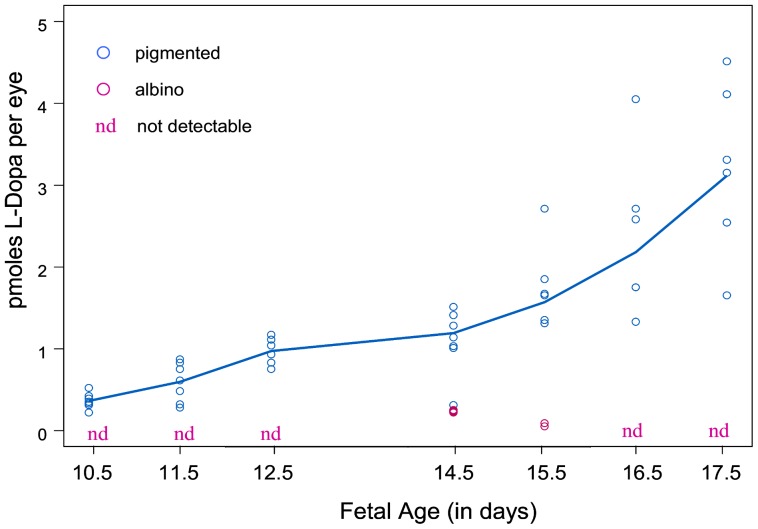
Figure 1. The Content of L-Dopa in Fetal Pigmented and Albino Eyes from Embryonic Day 10.5 to 17.5.
The L-Dopa content of individual eye samples appears on the graph according to fetal age. Fetal eyes were extracted for amine content at 7 ages ranging from E10.5, the day of first tyrosinase expression, to E17.5. Samples contained as few as the two eyes of a single fetus to as many as 12. L-Dopa was measured in every pigmented eye sample from E10.5 onward. No additional catecholamines were found. The quantity of L-Dopa in individual samples extracted from pigmented eyes is represented by the blue circles on the graph. Little or no L-Dopa was found in the extracts of albino eyes at any age (marked as “nd”, not detectable). The pink circles are the values from the three albino eye samples in which traces of L-Dopa were detectable. The solid blue line, obtained by non-parametric smoothing, reflects the temporal trend in the L-Dopa values for pigmented eyes. Also evident is the increased variability in L-Dopa content as the pigmented fetuses grow.

