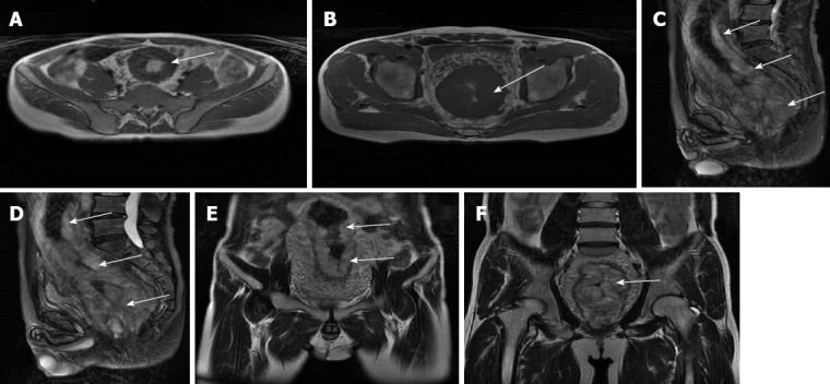
Figure 2.

Magnetic resonance imaging showed a significant thickness of the rectal wall, extending to the distal edge of the anus, with a narrow lumen (arrows). A, B: Horizontal plane imaging; C, D: Sagittal plane imaging; E, F: Coronal plane imaging.
