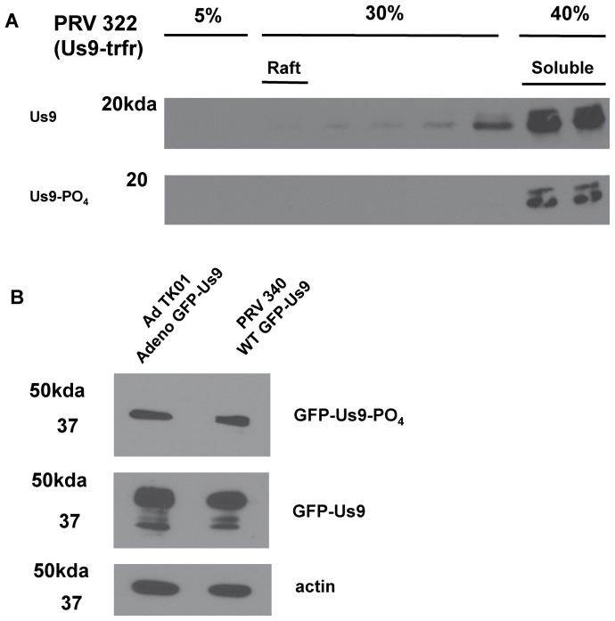
Figure 4. Molecular mechanisms of Us9 phosphorylation.
WB analysis with indicated antibodies probing samples from: A.) Differentiated PC12 cells 14 hours post-infection with PRV 322 followed by lipid raft float preparation. Samples were collected from a discontinuous 5%–30%–40% Optiprep gradient. DRMs localize to the 5%–30% interface, while solubilized membrane proteins remain at the 30%–40% interface. Each 1 mL fraction from this gradient was run and probed with polyclonal anti-Us9 antibody to detect total Us9 protein content and phospho-specific monoclonal antibody to detect only phosphorylated Us9. B.) Whole cell lysates of differentiated PC12 cells transduced with Ad TK101 for 24 hours.

