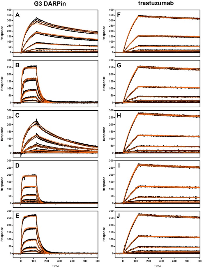
Figure 3. SPR binding sensorgrams for the interaction of HER2 wild type and mutant proteins with immobilized DARPin G3 (left panels) and trastuzumab (right panels).
Injected analyte (HER2) protein construct: wild type (A and F), Leu 525→Ala (B and G), Ser 551→Ala (C and H), Val 552→Ala (D and I) and Phe 555→Ala (E and J). Typically, injected HER 2 protein concentrations were diluted three-fold in running buffer from 81 nM to 1 nM except in B, D and E where the concentration series were diluted from 729 nM down to 9 nM. Overlayed triplicate binding responses are shown (black lines). Binding data were globally fit to a simple 1∶1 interaction model (orange lines).

