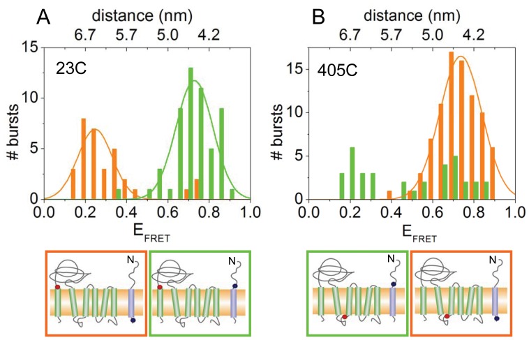
Figure 2. FRET histograms of YidC labeled at position 23 in the periplasmic region (A) and at position 405 in the cytoplasmic region (B).
Pf3 coat protein labeled at residue 16C (green bars) or 48C (red bars) was added, respectively. FRET efficiencies of individual bursts of Atto520-Pf3 protein with Atto647N-YidC and the resulting distance between the two probes were calculated. The positions of the Atto520 and Atto647N label are indicated by blue and red dots, respectively (lower panels).

