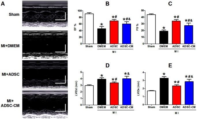
Figure 4. Cardiac function in MI mice after treatment with ADSC and ADSC-CM.
A: Representative M-mode images (long-axis view) of hearts with sham surgery and infracted hearts 4 weeks post-MI. B-C: Left ventricular ejection fraction (EF) and fraction shortening (FS) at 4 weeks post-MI. D-E: Diastole left ventricle internal diameter (LVIDd, mm) and systolic left ventricle internal diameter (LVIDs, mm) at 4 weeks post-MI (N = 7, * p<0.05 versus Sham; # p<0.05 versus MI+DMEM; & p<0.05 versus MI+ADSC).

