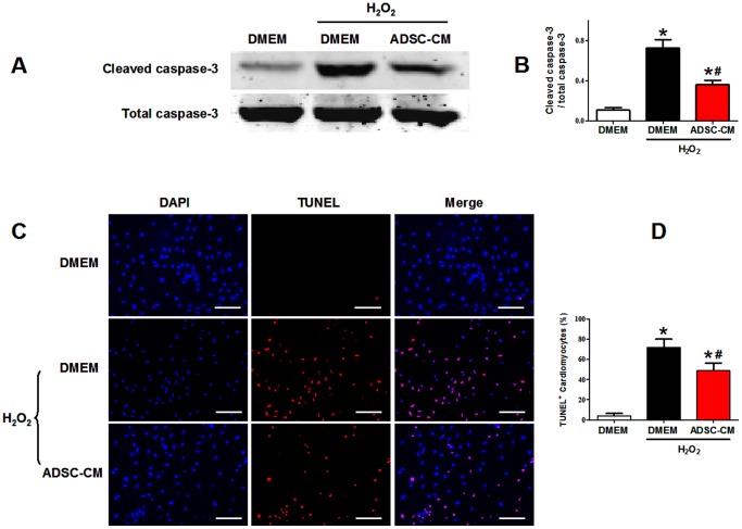
Figure 8. The antiapoptotic effect of ADSC-CM on cardiomyocytes subjected to oxidative stress.
A-B: Representative figures (A) and quantification (B) of western blotting analysis of cleaved caspase-3 of NRVMs subjected to H2O2 treatment (N = 5). C: Representative images of TUNEL labeling (TMR-red) and cell nuclei (DAPI-blue) of cardiomyocytes under H2O2 treatment. D: Quantification of TUNEL staining. TUNEL positive rate = (TUNEL-positive nuclei/DAPI-positive nuclei)×100% (N = 9). * p<0.05 versus control without H2O2; # p<0.05 versus control with H2O2. Scale bars represent 100 µm.

