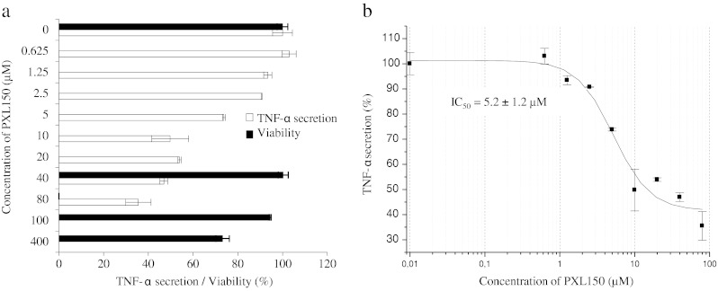
Fig. 3.

Effect of PXL150 on TNF-α secretion and viability of LPS-induced PMA-treated THP-1 cells. The peptide was added to cells in triplicate (n = 3) 30 min after the addition of LPS (0.1 ng/ml). Cytokine levels were measured in the cell supernatants by ELISA after 6 h of stimulation. a TNF-α secretion (white bars) is presented as relative secretion (%) ± SEM, with stimulated cytokine levels without peptide added set to 100 %. Viability of THP-1 cells (black bars) is presented as relative viability (%) ± SEM, with viability in stimulated cells without peptide set to 100 %. The cell viability was only measured for the peptide concentrations of 0, 40, 100 and 400 μM. b Data were fitted to the dose–response curve using a four-parameter fit in the Origin software. The IC50 value was automatically calculated by the software Origin
