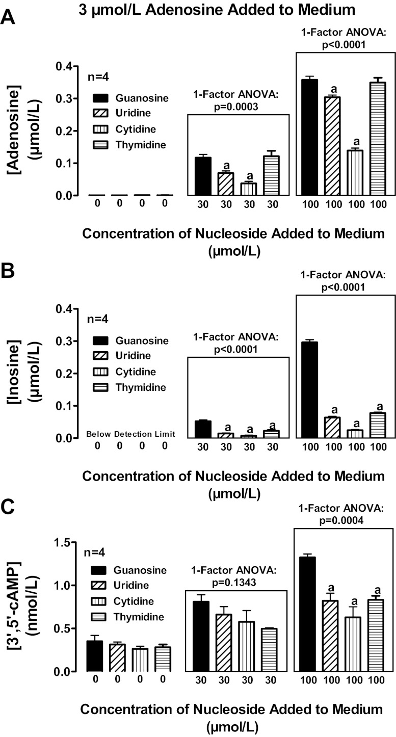
Fig. 12.
Rat preglomerular vascular smooth muscle cells were incubated for 1 h with various concentrations of guanosine, uridine, thymidine, or cytosine plus adenosine (3 μmol/l). The medium was assayed for adenosine (A), inosine (B), and 3′,5′-cAMP (C) by mass spectrometry. Values represent means ± SE. aSignificantly different from guanosine at the corresponding concentration.

