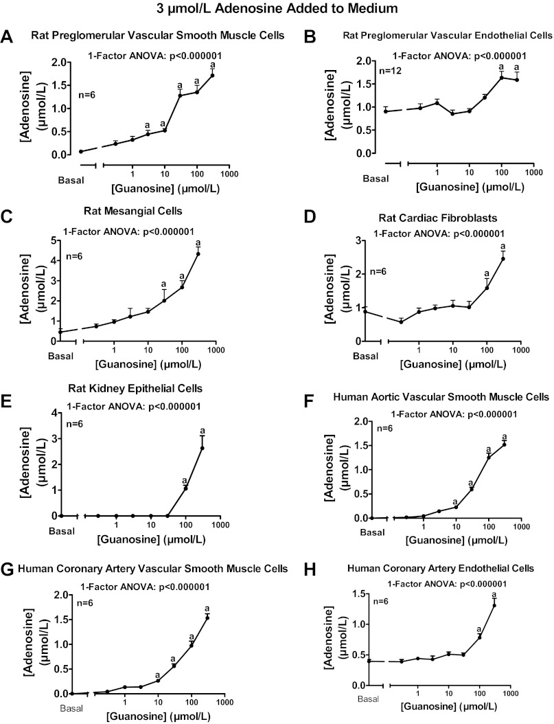
Fig. 7.
Rat preglomerular vascular smooth muscle cells (A), rat preglomerular vascular endothelial cells (B), rat mesangial cells (C), rat cardiac fibroblasts (D), rat kidney epithelial cells (E), human aortic vascular smooth muscle cells (F), human coronary artery vascular smooth muscle cells (G), and human coronary artery endothelial cells (H) were incubated for 1 h with various concentrations of guanosine [0 (basal), 0.3, 1, 3, 10, 30, 100, or 300 μmol/l] plus adenosine (3 μmol/l). The medium was assayed for adenosine by mass spectrometry. Values represent means ± SE. aSignificantly different from basal.

