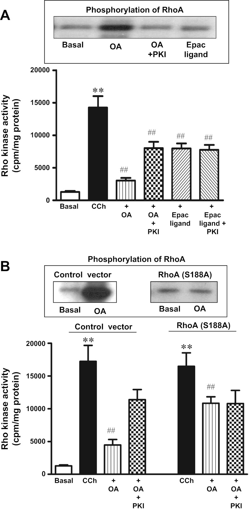
Fig. 4.
OA-induced phosphorylation of RhoA and inhibition of Rho kinase activity. A: RhoA phosphorylation (top). Freshly dispersed gastric smooth muscle cells labeled with 32P were incubated with OA (10 μM) in the presence or absence of myristoylated PKI (1 μM) or the cAMP analog that selectively activates Epac (8-pCPT-2′-O-Me-cAMP, 10 μM) for 10 min. RhoA immunoprecipitates were separated on SDS-PAGE, and RhoA phosphorylation was identified by autoradiography. In some experiments, cells were incubated with myristoylated PKI (1 μM). Rho kinase activity (bottom). Freshly dispersed muscle cells were treated with OA (10 μM) or 8-pCPT-2′-O-Me-cAMP (10 μM) in the presence or absence of myristoylated PKI (1 μM) for 10 min. Rho kinase activity was measured by immunokinase assay as described in materials and methods. Results are expressed as cpm/mg protein. Values are means ± SE of 4 experiments. **P < 0.01 vs. basal, ##P < 0.01 vs. CCh. B: RhoA phosphorylation (top). Cultured muscle transfected with control vector or vector containing phosphorylation-deficient RhoA mutant (S188A) were labeled with 32P and incubated with OA (10 μM) for 10 min. RhoA immunoprecipitates were separated on SDS-PAGE, and RhoA phosphorylation was identified by autoradiography. Rho kinase activity (bottom). Cultured muscle transfected with control vector or vector containing phosphorylation-deficient RhoA (S188A) were treated with CCh (0.1 μM) in the presence or absence of OA (10 μm) for 10 min. In some experiments cells were incubated with myristoylated PKI (1 μM). Rho kinase activity was measured by immunokinase assay. Results are expressed as cpm/mg protein. Values are means ± SE of 4 experiments. **P < 0.01 vs. basal. ##P < 0.01 vs. CCh.

