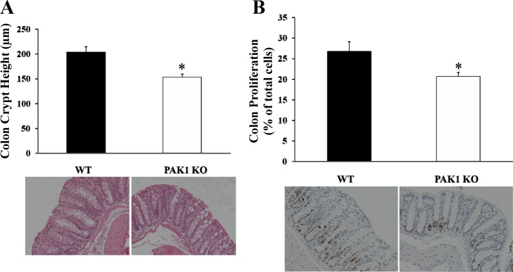
Fig. 4.

Proliferation in the colorectal mucosa was decreased in PAK1 KO mice. The colons from both WT and PAK1 KO mice were dissected, fixed, and stained as described in materials and methods. Colonic crypts were visualized by hematoxylin and eosin staining, and the crypt height was measured under the microscope at 20 times magnification (A). Proliferation was determined as the ratio of Ki67-stained cells to the total cells counted per field under the microscope (B). Results are summarized from 6–8 WT or PAK1 KO mice. *P < 0.05.
