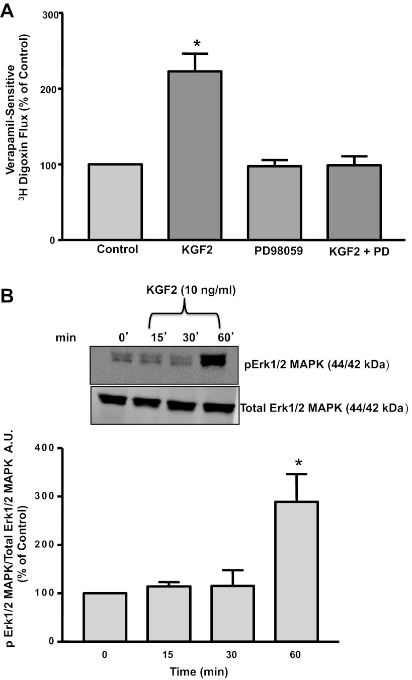
Fig. 3.
A: effect of Erk1/2 mitogen-activated protein kinase (MAPK) inhibitor on KGF2-induced stimulation of Pgp activity in Caco-2 cells. Postconfluent Caco-2 cells were pretreated with the specific inhibitor of Erk1/2 MAPK PD-98059 (30 μM) for 60 min in the serum-free cell culture medium and then coincubated with KGF2 (10 ng/ml) in serum-free cell culture medium supplemented with 0.2% BSA for 60 min. [3H]digoxin flux (1 μM) was measured as described in materials and methods. Results are expressed as %control and represent means ± SE of 5 separate experiments performed in triplicate. *P < 0.05 compared with untreated control. B: KGF2 induces Erk1/2 MAPK phosphorylation in Caco-2 cells. Caco-2 cells were incubated with KGF2 (10 ng/ml) in serum-free cell culture medium supplemented with 0.2% BSA for different time intervals ranging from 15 to 60 min. After the cells were washed with 1× PBS, extracted proteins (75 μg) were subjected to Western blot analysis on 12% SDS-polyacrylamide gel using phospho-specific Erk1/2 MAPK antibody (pErk1/2 MAPK). The blots were stripped and reprobed with the ERK1/2 MAPK antibody (total Erk1/2 MAPK) to indicate equal loading of protein in each lane. A representative blot of 3 different experiments is shown. The data were quantified by densitometric analysis and expressed as arbitrary units and represent means ± SE of 3 separate experiments. *P < 0.05 compared with untreated control (0 min).

