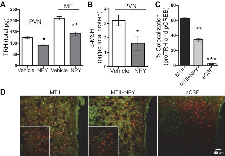
Fig. 5.
NPY reduced TRH and α-MSH in the PVN and blocked the MTII increase in pCREB in TRH neurons. A and B: male rats were infused icv with 5 μg NPY, and 60 min later PVN and ME samples were collected for RIA analyses. A: TRH peptide content in the PVN (n = 9/group, t-test, P < 0.05) and ME (n = 9/group, t-test, P < 0.05). Results were repeated for PVN TRH peptide content with the same result (control n = 4, NPY n = 4). B: α-MSH peptide content in the PVN (control n = 8, NPY n = 5, t-test, P = 0.03). C and D: male rats were infused icv with 5 μg NPY and/or 5 μg MTII. Tissue from the PVN was subjected to double IHC for cytoplasmic pro-TRH (red fluorescent) and nuclear pCREB (green fluorescent); 36 fields per section and condition were analyzed. C: colocalization between pro-TRH and pCREB in rats treated with aCSF, MTII, or MTII + NPY (n = 9/group, ANOVA, P < 0.0001). D: representative low and high (bottom left) magnification images are shown for each treatment condition. Rat PVN tissue was treated with aCSF, MTII, or MTII + NPY. Tukey's test was used for all post hoc analyses. See text for definitions. Data are means ± SE. *P < 0.05, **P < 0.01, ***P < 0.001 vs. control.

