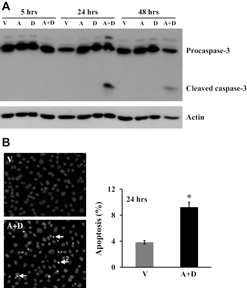
Fig. 2.
Effect of sustained adenosine exposure on cultured lung EC apoptosis. A: bovine pulmonary artery ECs (PAEC) were incubated with vehicle (V) or 50 μM adenosine (A) in the absence or presence of 50 μM deoxycoformicin (D) for indicated times. Apoptosis was assessed by the proapoptotic, cleaved form of caspase-3. The same immunoblots were stripped and reprobed for actin to control for protein loading. Data represent three independent experiments. B: bovine PAEC were incubated with V or 50 μM A plus 50 μM D for 24 h, and apoptosis was assessed by 4,6-diamidino-2-phenylindole (DAPI) staining of apoptotic nuclei. Representative images from five independent experiments are shown. Arrows indicate apoptotic nuclei. Data are means ± SE, expressed as the ratio of apoptotic cells to the total counted cells × 100 (%). About 600 cells in three high-power fields for each group in each independent experiment were analyzed by a blinded observer. *P < 0.05 vs. V-treated EC.

