
Fig. 9.
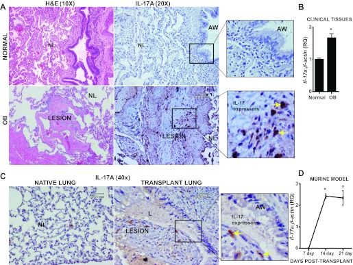
IL-17A is expressed in clinical OB lesions after lung transplant. A: immunohistochemical staining showing expression of IL-17A in a representative human subject with OB and normal lungs. Hematoxylin and eosin staining reveals the respective lung architecture. Inset in OB: IL-17 expressing cells in the fibrotic plugs. Inset in normal lung: epithelial and subepithelial tissues but no staining for IL-17. Representative lesions are presented subsequent to examining lung biopsies from 3 different patients. B: mRNA expression of IL-17A normalized to β-actin in human lung tissues. Values represent mean ± SE; *P < 0.05; normal = 4, OB = 3. C: immunohistochemical staining showing expression of IL-17A in a representative mouse orthotopic allograft model in both native (right) and allograft (left) lungs. Transplant lung shows IL-17 expressing cells in the fibrotic plugs (yellow arrows). Native lung shows epithelial and subepithelial tissues but no staining for IL-17. Representative lesions are presented subsequent to examining lung biopsies from 3 different mice. D: mRNA expression of IL-17A normalized to β-actin in mouse orthotopic lung allograft model at days 7, 14, and 21 days after lung transplant. Values represent means ± SE; *P < 0.05; n = 4.