
Fig. 3.
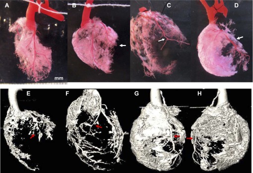
Fibrosis in remote/nonischemic myocardium. Top row: regular photos. A: control. B: LCA ligation 1 mo (MI-1m). C: ischemia/reperfusion injury 1 mo (I/R-1m) with residential vessel tail distal to ligated site. D: Ab+I/R+DeAb. Bottom row: micro CT images. E: MI-1m. F: I/R-1m. G: anterior view of Ab+I/R+DeAb. H: posterior view of Ab+I/R+DeAb. Arrow: ligated site.